")
")
Scanning Electron Microscope, SEM
Laboratory equipment is purchased as part of the project "Research Infrastructure for Campus-based Laboratories at the University of Rijeka" (RISK), which is co-financed by the European Union under the European Regional Development Fund.
![]()
Laboratory: O-120
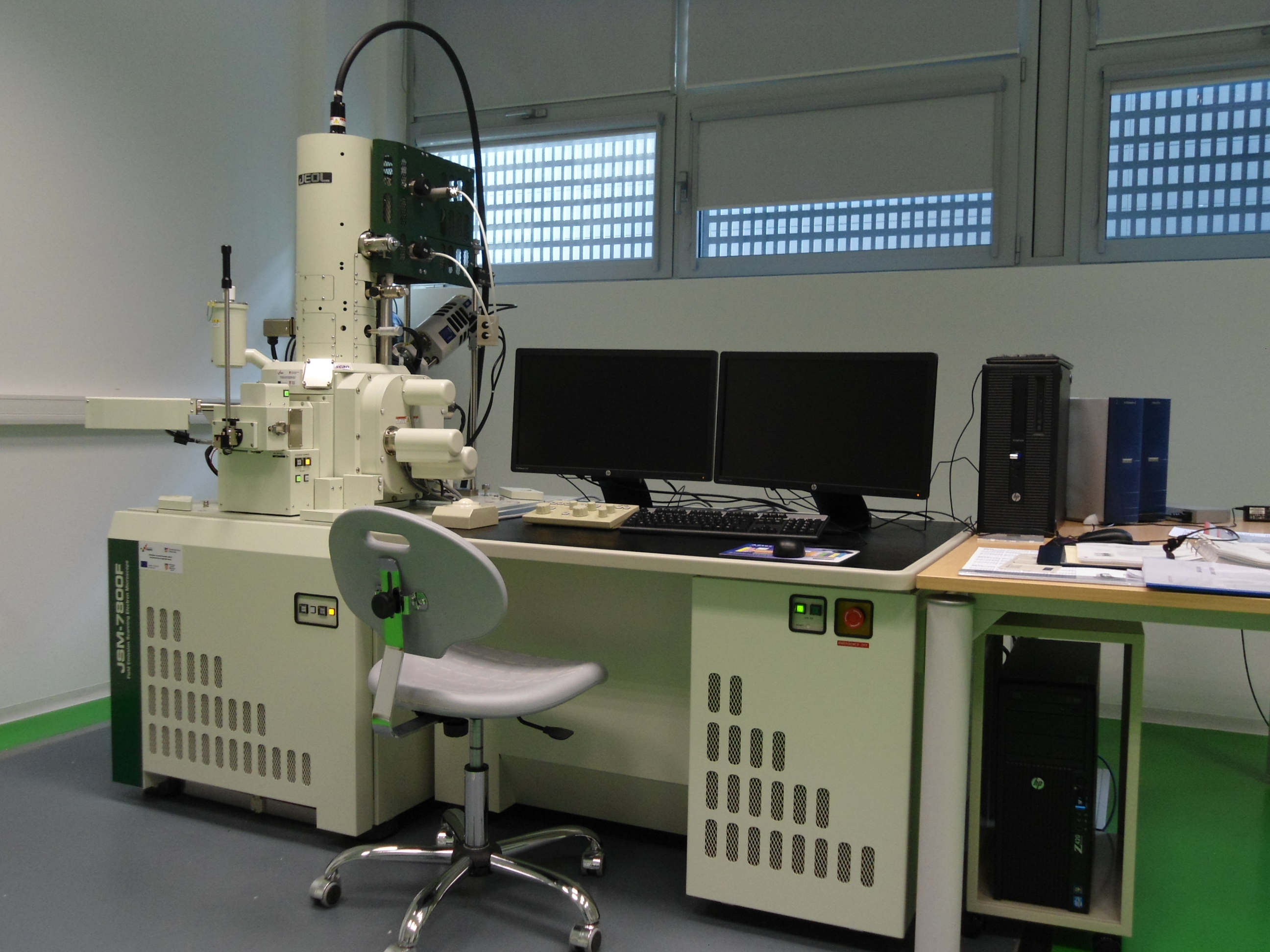
JSM-7800F Field Emission Scanning Electron Microscope
| Short description: |
Scanning Electron Microscope (SEM): a type of electron microscope that permits the observation and characterization of heterogeneous organic and inorganic materials on a nanometre (nm) to micrometre (μm) scale. |
| Main purpose: |
The SEM is capable of obtaining 3D-like images of the surfaces of a very wide range of materials. The surface area to be examined or the micro-volume to be analysed is irradiated with a finely focused electron beam, which may be swept in a raster across the surface of the specimento form images or may be static to obtain analysesat one point. Signals produced from the interaction of the electron beam with the sample are in the form of secondary electrons (emitted from the sample), backscattered electrons (from the impinging electron beam) or characteristic X-rays. Signals are obtained from specific emission volumes within the sample and can be used to examine many characteristics of the sample (surface topography, crystallography, chemical composition, etc.). |
| Technical specifications: |
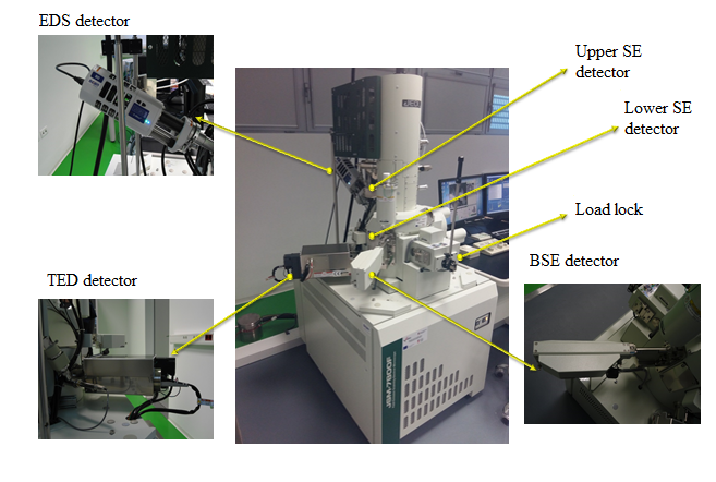
The SEM is a JEOL Field Emission Scanning Electron Microscope (JSM-7800F) with maximal resolution of 0.8 nm, acceleration voltage of 0.01 – 30 kV and the magnification range: x25 – x1000000. It is equipped with the following detectors:
|
| Year of manufacture: |
2014 |
| Source of founding: |
„Research Infrastructure for Campus-based Laboratories at the University of Rijeka“ project financed by ERDF |
| Contacts: |
dr. Ivna Kavre Piltaver (+ 385 51 584 618, ivna.kavre@uniri.hr) |
Contact
FACULTY OF PHYSICS
UNIVERSITY OF RIJEKA
Ulica Radmile Matejčić 2
51000 Rijeka
Tel.: +385 51 584 600
Fax: +385 51 584 649
Email: fizika@phy.uniri.hr
 |
 |
|
|
|
 |
 |
 |
|
 |
|
|
|
|
 |
|
 |
|
 |
|
 |
|
 |
 |